DIFFERENTIAL DIAGNOSIS IN CLINICAL SURGERY
Patchy gangrene of the skin
1.Vasculitis.
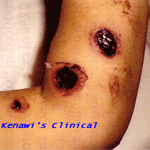
Patchy gangrene due to vasculitis
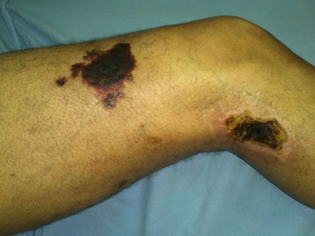
2.Ecthyma Gangrenosum.
Classically described as occurring in Pseudomonas aeruginosa bacteraemia, but can be caused by other organisms. It also affects other immunocompromised patients as those with severe malnutrition, diabetes, etc. It starts as well-circumscribed oedematous lesions (vesicles) that quickly acquire a haemorrhagic centre and an erythematous periphery. Increase in size is associated with central ulcerative necrosis spreading towards the periphery of the lesion (Fig. 1).
Gangrenous Ecthyma
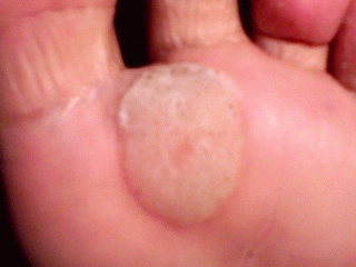
Ulcers on the sole of the foot raised above skin level
1.Squamous cell carcinoma (Epithelioma).
The edge is raised and everted and at a higher level than the floor.
2.Flat warts (Verruca plana).
May be simultaneously present in other sites of the body. It affects multiple sites in immunocompromised patients as those with severe malnutrition, diabetes, etc. (Click here to view multiple affection).
Flat wart (Verruca plana) of the left foot
Discoloration and associated thickness of the nipple and areola
1.Adenomatosis of the nipple.
2.Early (non-ulcerated) Paget's disease of the nipple.
This cannot be differentiated clinically from adenomatosis of the nipple except by highly experienced surgeons. Immunophenotyping must be done on a biopsy specimen. Paraffin sections are to be treated with monoclonal antibodies against EMA and S100 using streptavidin biotin peroxidase system. If the sections treated for EMA reveal positive cytoplasmic staining of the cells by chromogen and those treated for S100 reveal no such staining, then this is early Paget's disease of the Nipple.
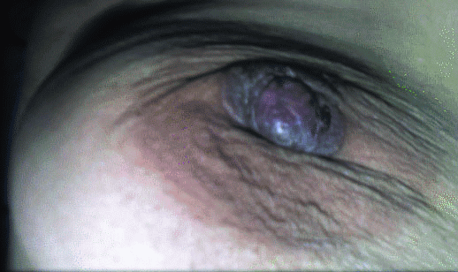
Early Paget's disease of the Nipple
Warty lesions of the scalp and skin
1.Warts.
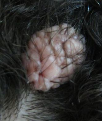
2.Syringocystadenoma Papilliferum.
Is often present at birth and most often presents on the scalp as a warty nodule (Fig. 4). It frequently arises in association with a naevus, such as naevus sebaceus. It behaves in a benign fashion. Histologic sections show an exophytic/endophytic lesion creating a cystic space with a connection to the surface.The bulbous projections are loaded with plasma cells. The epidermis is papillomatous and acanthotic.
Syringocystadenoma Papilliferum of the scalp
![]()
Students' & Continuing Surgical Education
